
-
Email support@jcmcri.org
-
Address Boduppal, Hyderabad, India.
Grodno state Medical University, Belarus.
*Corresponding author: Bon LI
Candidate of biological science, Assistant professor
of pathophysiology department named D. A.
Maslakov, Grodno State Medical University; Grodno
State Medical University, 80 Gorky St,230009,
Grodno, Belarus.
Email: asphodela@list.ru
Received: July 16, 2026
Accepted: July 22, 2026
Published Online: July 23, 2026
Journal: Journal of Clinical, Medical Case Reports and Images
Copyright: © Bon LI (2026). This Article is distributed under the terms of Creative Commons Attribution 4.0 International License
Citation: Bon EI, Sidzineuskaya HI, Sorokina TS, Malenovskaya MY, Koval A, Dobrinets L. Biochemical changes in muscle tissue during compression trauma induced by hemostatic tourniquet application. J Cli Med Case Rep Images. 2026; 1(1): 1002
Keywords: Hemostatic tourniquet; compression trauma; muscle tissue, ischemia; biochemical changes; total nitrogen, actomyosin complex; creatine phosphate; ATP; glycogen; lactic acid; respiratory quotient; proteolysis; enzyme activity (dehydrogenases, ATPase); muscle edema; reperfusion; recovery period; irreversible disorders; amytal sleep.
The study of biochemical processes in muscle tissue during traumatic injuries and compression is of key importance for clinical, particularly surgical, practice. One common but insufficiently studied type of impact is the application of a hemostatic tourniquet — a method of temporary bleeding control widely used in military field surgery and continuing to be employed in modern operative surgery. Despite the regulated time limits (no more than 2–2.5 hours), in real-world conditions, especially wartime, these restrictions are often violated, leading to profound pathological changes in tissues. This review, based on experimental data, analyzes the nature and extent of biochemical shifts in muscles depending on the duration of tourniquet compression and traces the features of the recovery period after its removal.
In a number of studies conducted mainly on artificial systems (muscle tissue homogenates, extracts, protein solutions, etc.), significant changes in the properties of muscle proteins were found under the influence of penetrating radiation [1, 2]. Simply. In these studies, huge doses of penetrating radiation were used, in some cases affecting for a very long time.
The study of biochemical changes in traumatic muscle injuries is undoubtedly of great interest for clinical and, in particular, surgical practice [3]. Unfortunately, most of the issues in this area are relatively poorly covered in the specialized biochemical literature [4]. The biochemical processes in muscles during the application of a hemostatic tourniquet, which can be considered as a form of compression trauma, have been studied in much greater detail [5]. The application of an elastic bandage or tourniquet is a common method of temporarily stopping bleeding in limb injuries [6]. According to G. F. Nikolaev, during the Great Patriotic War, the application of a standard or improvised tourniquet was used in more than 65% of cases to temporarily stop bleeding [7]. The application of a hemostatic tourniquet is also used in clinical practice during various surgeries on the limbs [8]. It is generally accepted that the time of application of a tourniquet should not exceed 2-2.5 hours [9]. In peacetime, compliance with these time limits is usually feasible, but in wartime, it may not always be possible [10]. According to the analysis of materials from the Great Patriotic War, compliance with the two-hour time limit was achieved in only 46.2% of cases, which is less than half [11]. In 12.8% of cases, the tourniquet was applied for more than six hours. Therefore, studying the nature and extent of pathological changes and the depth of biochemical shifts that occur in tissues depending on the duration of the tourniquet application is a crucial task [12]. It is important to note that the application of a tourniquet is a complex form of compression that causes a range of harmful effects [13]. These effects include complete or partial denervation, circulatory disorders, general and local intoxication, and more. The extent of biochemical changes in muscle tissue depends on various factors, such as the time, location, and degree of compression, the thickness of the subcutaneous fat layer and muscles, the ambient temperature, and more.
A detailed study of the metabolism in muscles after applying a hemostatic tourniquet was conducted [14, 15]. The described biochemical changes were primarily observed in animal experiments (on rabbits and dogs), and in most cases, the results of similar measurements in the symmetrical muscles of the intact limb were used for comparative evaluation of the data obtained after applying the tourniquet. This is important because the intact limb often exhibits similar changes to those observed in the limb that has been tied with a hemostatic tourniquet.
The total nitrogen content in the muscles below the tourniquet site does not change until the tourniquet is removed, regardless of the duration of compression, which can range from one hour to a day [16]. After the tourniquet is removed after three hours and blood circulation is restored, the total nitrogen content decreases slightly in relation to the weight of the raw tissue, which is likely due to the development of edema [17].
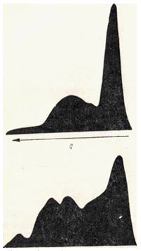
With longer compression (6 hours), the amount of total nitrogen in the muscle tissue and in the extracts extracted with high-ionic-strength saline solutions and, to a lesser extent, with low-ionic-strength saline solutions, decreases even more (Table 22) [18The electrophoretic pattern of protein extracts obtained from the muscles of animals killed 24 hours after 6 hours of tourniquet application indicates certain shifts in the protein composition of the damaged muscles and, in particular, a relative decrease in the proteins of the actomyosin complex and an increase in the myoalbumin fraction (Fig. 21) [19]. However, it is possible that prolonged tourniquet application leads to partial denaturation of muscle proteins, which may affect the protein composition of the extracts.
| Muscle | мг | Extract with Weber's solution1 | Phosphate extract. The pH of the buffer is 7.8 (10.1) | ||
|---|---|---|---|---|---|
| мг | % | мг | % | ||
| Control | 32 | 18 | 56,2 | 7,2 | 22,5 |
| The tourniquet | 28 | 13,8 | 45,7 | 6 | 21,4 |
(in mg per 1 g of raw tissue and as a percentage of total nitrogen), according to V. A. Yuryev
A decrease in the content of myosin and water-soluble proteins in muscles after the application of a tourniquet was noted [20]. The content of non-protein nitrogenous products in damaged muscle tissue also changes.
The concentration of residual nitrogen increases in the first three hours after the application of the tourniquet, but then decreases. The amount of amine nitrogen increases significantly, reaching a maximum by the sixth hour of compression. During this period, the amount of amine nitrogen in the muscle tissue of the damaged limb is one and a half times higher than in the control limb of the animal.
The application of a tourniquet also increases the content of ammonia in muscles, and prolonged compression increases the content of amide nitrogen. The amount of creatine in muscles decreases significantly after compression. The increase in the content of low-molecular-weight nitrogenous products after applying a tourniquet appears to be associated with an increase in proteolytic processes in damaged muscle tissue.
Among the most common indicators of metabolic disorders in muscle tissue, the most striking are the decrease in respiration and the change in the respiratory quotient [1]. While the respiratory quotient in the intact limb is approximately 1, it drops to 0.7-0.6 in the muscles of the injured limb [2]. This indicates that under the changed conditions, carbohydrate oxidation is disrupted, and other organic substances become the substrate for respiration [3]. However, the content of glycogen in the muscles also decreases significantly. After 3 hours of compression, the glycogen content in the muscle is approximately 80% of the initial value, and after 8 hours, it is approximately 20%. By the end of the day, glycogen in the muscle tissue has completely disappeared. In parallel with the decrease in glycogen, the concentration of lactic acid increases [4, 5].
Significant changes also occur in the content of phosphorus compounds [6]. Creatine phosphate disappears from the muscle tissue most rapidly, with most of it being consumed within the first hour. The content of ATP does not change during the first two hours, but it drops sharply (by 40%) by the end of the third hour and completely disappears by the eighth hour of compression [7]. Simultaneously with the decrease in creatine phosphate and ATP, the amount of inorganic phosphate increases [8]. The stability of ATP content during the first hours, particularly when creatine phosphate is almost completely absent in the muscle tissue, is noteworthy. This is attributed partly to the ability to maintain ATP levels through glycolysis, but primarily to the decrease in ATPase activity in the muscle tissue [9]. However, the significant decrease in ATP content despite the presence of significant glycogen reserves in the muscle tissue suggests a different explanation [10]. The first assumption is contradicted by a noticeable decrease in ATP content, despite the presence of significant glycogen reserves in muscle tissue.
The activity of various muscle enzymes, both hydrolytic and oxidative, undergoes significant changes [11]. It is noteworthy that the activation of muscle enzymes such as amylase and maltase occurs simultaneously with the inhibition of ATPase activity [12].
At the same time, there is evidence that, along with the inhibitory effect of compression on the ATPase activity of myosin, there is an activation of water-soluble muscle ATPase [13]. The initial period of some activation is also characteristic of glycerophosphate dehydrogenase [14]. The activity of lactate dehydrogenase remains relatively stable for the first 6 hours, but it subsequently decreases [15]. The activity of succinate dehydrogenase remains elevated for an even longer period [16]. The glucose content in the muscle decreases slightly in the first hours and then remains relatively stable [17].
The ability of muscle tissue to dehydrogenate glucose under the conditions of the Thunberg experiment (with methylene blue) increases significantly during the first three hours of the tourniquet [18]. Over the following period, it gradually decreases and disappears after 12 hours [19].
Other changes that occur in the muscles after the application of a tourniquet include a decrease in the total content of lipids, an increase in the content of water (edema), and a decrease in the pH value to 5-5.5 [20].
It is of significant interest to study the biochemical changes in the muscle after the application of a tourniquet in the absence of the central nervous system.
The application of a tourniquet in animals that are in a state of amytal sleep is characterized by some differences from the described pattern of metabolic disorders. When the central nervous system is disrupted, the dehydration of succinate and lactate is significantly activated, and the overall reduction value increases. At the same time, the disruption of the central nervous system eliminates the activation of certain other oxidative enzymes that usually occur under the influence of compression. According to these studies, the adaptive reactions that the muscle can exhibit in the first period after the application of a tourniquet are possible only due to the influence of the central nervous system.
The data on the changes in the activity of several enzymes indicate a certain restructuring of metabolic processes, which is to some extent compensatory in nature, especially noticeable in the first hours after the application of a tourniquet. In particular, this restructuring involves an increase in anaerobic processes in the muscle under conditions of oxygen deprivation. Similar changes in the nature of metabolic processes, which are a manifestation of adaptation to changed conditions, are known to occur in physiological conditions of the muscular system. A vast amount of experimental data in this area is presented in numerous studies. However, it would be incorrect to assume that all the biochemical changes that occur in the muscles after the application of a tourniquet are compensatory in nature. A significant portion of the detected changes is undoubtedly a manifestation of pathologically altered metabolic processes that worsen the functional state of the muscles.
Significant changes occur in the muscle after the blood stopping tourniquet is removed, and the degree and completeness of the normalization of biochemical processes in the muscle tissue depend entirely on the time of the tourniquet application [1]. In the first 2 hours after the tourniquet, which was applied to the limb for 3 hours, the oxygen consumption of the muscle tissue increases significantly. The respiratory coefficient decreases to 0.35. At the same time, there is a significant increase in respiration in the intact limb, accompanied by a slight decrease in the respiratory coefficient. The amount of glycogen increases rapidly, and within 1 to 6 hours, the glycogen content in the muscles of the compressed limb is higher than in the intact limb. This high glycogen content persists for about 24 hours, after which it returns to its original level.
The amount of lipids in the muscle decreases significantly after the tourniquet is removed, but then increases dramatically, and after 24 hours, the amount of lipids in the altered muscle is higher than the original level. It is interesting to note that if the tourniquet was applied to the limb during the animal’s amytal sleep, there is no significant increase in glycogen levels in the damaged muscles.
The level of phosphocreatine also recovers quite quickly after the tourniquet is removed, despite the fact that its content was close to zero before the tourniquet was removed. After the tourniquet is removed, the level of ATP initially continues to decrease, but then gradually increases, reaching its original level approximately 24 hours later [2].
The maximum changes are observed 20 hours after the tourniquet is removed. Rapid changes also occur in the content of non-protein nitrogenous products. The residual nitrogen in the muscles decreases while its amount in the blood increases. The content of amine nitrogen and ammonia in the muscles also decreases. After 24 hours, the content of these components in the muscle tissue returns to normal.
Issekutz, Hetenyi, and Winter studied the metabolism in the muscles of a dog after a 4-hour tourniquet was applied to the limb [3]. They observed similar phenomena in terms of oxygen consumption and ATP content. They identified three phases in the observed changes: the first phase, which lasted 40-60 minutes after the limb was freed from the tourniquet, was characterized by increased oxygen consumption by the muscles, a sharp increase in ATP content, and increased release of inorganic phosphorus into the blood. The second phase, which lasted 1.5-2 hours, was marked by the normalization of oxygen consumption, an increase in the release of lactic acid, and continued increased release of inorganic phosphorus into the blood, as well as a low content of ATP in the muscles. The subsequent third phase was characterized by a gradual normalization of metabolism in the altered muscles.
Jordan and Gray applied a tourniquet to the leg of rats for 4 hours and used ion-exchange chromatography to determine the ATP content at various time points [4]. The ATP content in the muscles of the leg that was tied with a tourniquet was reduced by approximately 10 times and remained at a relatively low level for some time after the tourniquet was removed.
After the tourniquet was removed, the pH of the muscle tissue increased to 7.4-7.6, while the reserve alkalinity of the blood decreased dramatically to 25-30. The amount of inorganic phosphorus and potassium in the muscles decreased, while the amount of chlorine and calcium increased. In the blood, the opposite was observed, i.e., the amount of phosphorus and potassium increased, while the amount of chlorine and calcium decreased [5]. According to MacPhee, after the tourniquet was removed, there was a sharp decrease in the concentration of potassium and an increase in the concentration of sodium in the muscles [6]. It is of some interest to note the parallelism between the degree of acidosis and the accumulation of amino acids in the muscles. According to Eckel, Pope, and Norris, a decrease in the potassium content in muscle tissue is accompanied by regular increase in the concentration of essential amino acids, in particularly lysine [7]. It is possible that the involvement of amino acids in maintaining an active reaction in the muscles is a reflection of their broader role in regulating the acid-base balance in the body.
The recovery of enzyme activity is relatively slow, and in the first hours after the tourniquet is removed, there is a tendency for some enzymes to continue to decrease in activity. This is particularly evident in enzymes such as lactate dehydrogenase and glucose dehydrogenase, which have zero activity three hours after the tourniquet is removed. Other enzymes exhibit activity levels ranging from 73% to 82% of their initial levels.
Succinate dehydrogenase exhibits relatively high resistance, and its activity returns to normal on the 5th day, while the activity of other enzymes returns to normal much later (after 10 days) [8].
It should be noted that the well-known surgical technique of short-term compression interruption has a positive effect [9]. Restoring blood circulation by removing the tourniquet for 5 minutes during a total of 4 hours of tourniquet application has a much less significant impact on the activity of several enzymes compared to continuous 4-hour compression [10].
The administration of small doses of ATP during the initial stage of recovery (ATP was administered subcutaneously to rabbits at a dose of 10-25 mg) significantly accelerated the normalization of the observed metabolic disorders [11].
The recovery period after removing a tourniquet that had been applied to the limb for 6 hours is significantly slower and less complete. The level of glycogen remains reduced for up to 47 days after the tourniquet is removed. By this time, the activity of dehydrogenases and ATPase is also significantly reduced [12].
Based on these experimental findings, it can be concluded that a 3- hour tourniquet application is the maximum duration during which the biochemical changes can be fully reversible [13]. After this duration, persistent functional and morphological disorders occur [14].
The presented analysis of experimental data shows that the application of a hemostatic tourniquet induces a complex set of biochemical disturbances in muscle tissue: from depletion of glycogen, creatine phosphate, and ATP stores to alterations in the activity of key enzymes and the accumulation of low molecular-weight nitrogenous products. It was revealed that during the first hours of compression, compensatory mechanisms (particularly anaerobic glycolysis) are activated in the muscles; however, as ischemia time increases, pathological changes progress. The critical threshold for the reversibility of these processes is 3 hours of tourniquet application: beyond this duration, the recovery of metabolism, enzyme activity, and tissue structure becomes incomplete and prolonged, and with 6-hour compression, persistent functional and morphological disorders develop. These findings underscore the need for strict monitoring of tourniquet application time and justify the search for methods (e.g., brief interruption of compression, ATP administration) capable of accelerating metabolic normalization in the post-ischemic period.